 |
|
 |
 |
 |
 |


Statins do not affect viability and maturation of pDCs. (A-C)
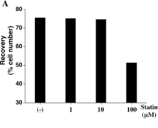
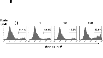
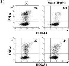
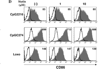
pDCs were cultured with 5 µM CpG 2216 in the absence or presence of different concentrations of simvastatin, as indicated in figures. After 24 h, viable cells were measured by a trypan-blue exclusion test (A) and annexin V staining (B). Percentages of annexin V-positive cells are indicated. (C) After 8 h of stimulation with CpG 2216, intracellular cytokine (IFN-α and TNF-α) production and surface BDCA4 expression by activated pDCs was analyzed by flow cytometry. Percentages of the cytokine-producing pDCs are indicated in each dot-blot profile. (D) CD86 expression on pDCs stimulated for 24 h with 5 µM CpG 2216, 5 µM CpG c274, or 100 µM loxoribine were analyzed by flow cytometry. The staining profiles of CD86 and isotype-matched control are indicated by shaded and open areas, respectively. Numbers in the histograms indicate the mean fluorescence intensity (MFI), which is calculated by the subtraction of MFI with the isotype control from that with CD86 mAb. Similar results were observed in three independent donors and the results of a representative experiment are shown.
pDCs were cultured with 5 µM CpG 2216 in the absence or presence of different concentrations of simvastatin, as indicated in figures. After 24 h, viable cells were measured by a trypan-blue exclusion test (A) and annexin V staining (B). Percentages of annexin V-positive cells are indicated. (C) After 8 h of stimulation with CpG 2216, intracellular cytokine (IFN-α and TNF-α) production and surface BDCA4 expression by activated pDCs was analyzed by flow cytometry. Percentages of the cytokine-producing pDCs are indicated in each dot-blot profile. (D) CD86 expression on pDCs stimulated for 24 h with 5 µM CpG 2216, 5 µM CpG c274, or 100 µM loxoribine were analyzed by flow cytometry. The staining profiles of CD86 and isotype-matched control are indicated by shaded and open areas, respectively. Numbers in the histograms indicate the mean fluorescence intensity (MFI), which is calculated by the subtraction of MFI with the isotype control from that with CD86 mAb. Similar results were observed in three independent donors and the results of a representative experiment are shown.


